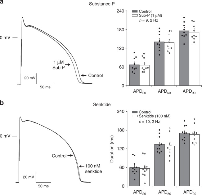
Fig. 7.
Effect of NK-3 receptor stimulation on action potentials of ventricular myocytes. (a–b, left panel) Ventricular APs elicited at 2 Hz under control conditions (Control) and in the presence of 1 µM Sub-P a or 100 nM Senktide b. (a–b, right panel) Bar graph showing average values for AP duration at 20, 50 and 90% of repolarization (APD20, APD50 and APD90) before (Control) and after application of 1 µM Sub-P (a, N = 5, n = 9) or 100 nM Senktide (b, N = 5, n = 10). Paired t-test. All values shown are mean ± SEM. *P < 0.05

