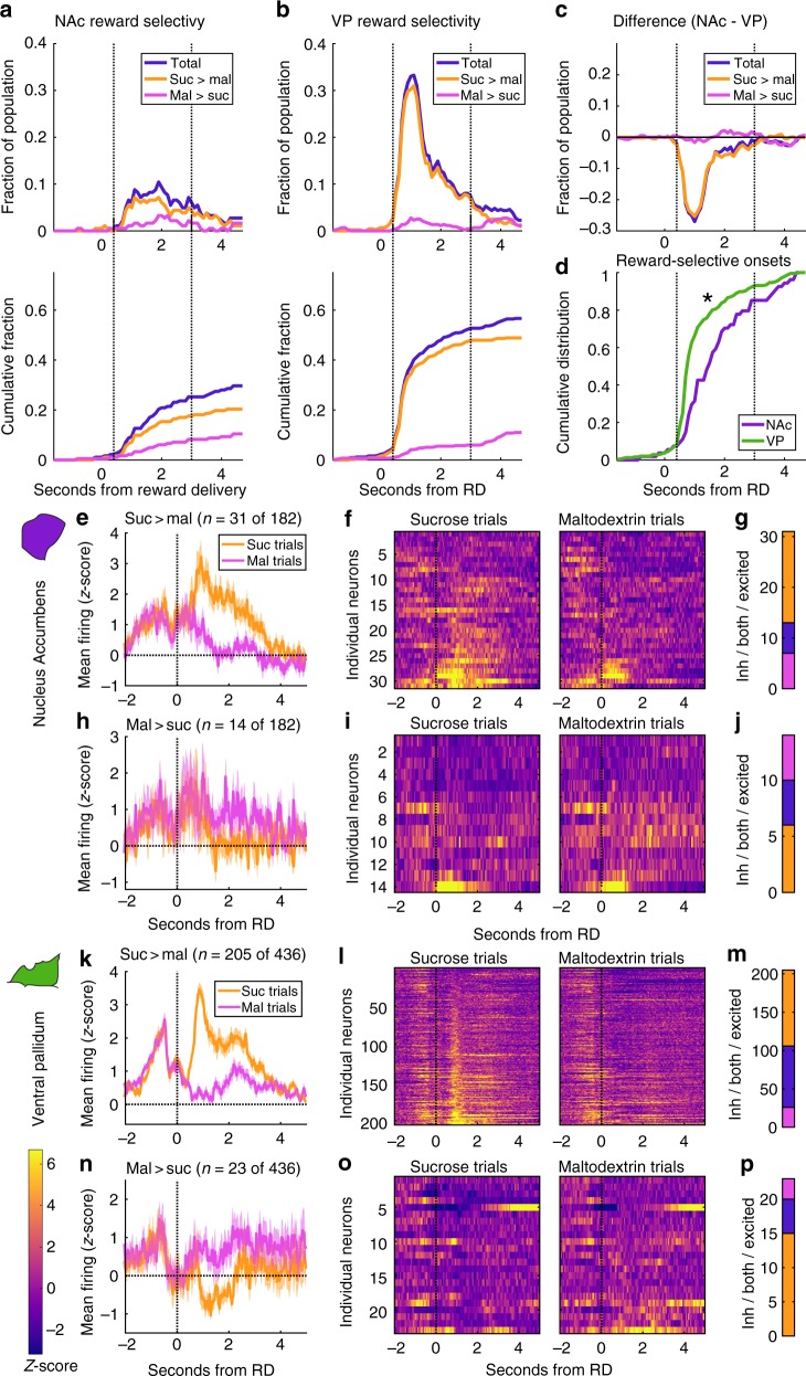
Fig. 2.
More neurons in VP fire selectively for sucrose and maltodextrin than in NAc. a, b Top panel: fraction of NAc (a) and VP (b) neurons meeting criteria for reward selectivity as a function of time after reward delivery. Plotted are total fraction of reward-selective neurons (blue) and, of those, neurons with greater firing for sucrose (orange) and greater firing for maltodextrin (pink). Bottom panel: Cumulative distribution of reward selectivity over time after reward delivery. c Subtraction of VP reward selectivity from NAc in each bin. Negative values indicate more selectivity in VP. d Cumulative distribution of reward selectivity onsets as a fraction of total reward-selective neurons. Asterisk indicates significantly earlier onsets in VP (F(1,290) = 12.7, p = 0.00071). e–g Neurons with greater firing for sucrose in any bin centered at 0.4–3 s. e Mean normalized firing rate for sucrose-selective neurons on sucrose (orange) and maltodextrin (pink) trials. Shading is SEM. f Heat maps of the normalized activity of individual sucrose-selective neurons on sucrose and maltodextrin trials. g Number of neurons with maltodextrin inhibitions (pink), sucrose excitations (orange), or both (blue). h–j Neurons in NAc with greater firing rate for maltodextrin in any of the bins centered 0.4–3 s. h Mean normalized firing rate for maltodextrin-selective neurons on sucrose (orange) and maltodextrin (pink) trials. Shading is SEM. i Heat maps of the normalized activity of individual maltodextrin-selective neurons in NAc on sucrose and maltodextrin trials. j Number of neurons in NAc with maltodextrin inhibitions (pink), sucrose excitations (orange), or both (blue). k–p Sucrose- (k–m) and maltodextrin- (n–p) selective neurons in VP, plotted as for NAc neurons in e–j

