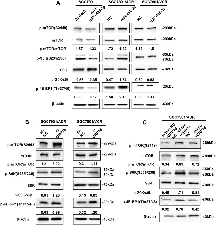
Fig. 6. miR-495-3p inhibits autophagy by activating mTOR signaling.
a Western blot analysis of phosphorylated mTOR (p-mTOR) and its main substrates 4E-BP1 (p-4E-BP1) and S6 (p-S6) in SGC7901 transfected with anti-miR-495-3p and GC MDR cells transfected with miR-495-3p. b The phosphorylation status of mTOR, S6K (p-S6K), and 4E-BP1 (p-4E-BP1) transfected with si-GRP78 and si-NC were measured by western blotting (c). Western blotting determines the phosphorylation of mTOR, S6K (p-S6K), and 4E-BP1 (p-4E-BP1) in SGC7901/ADR co-transfected with GRP78 3′-UTR-mutant overexpression vector/nc and miR-495-3p/nc. All values expressed as mean ± SD, n = 3 for each group. *P < 0.05, **P < 0.01, ***P < 0.001

