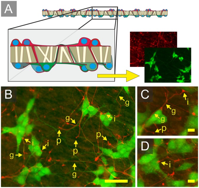
Figure 7.
Neurite growth of spatially separated SH-SY5Y neural populations across a 1.2 µM membrane. (A) Schematic demonstrating the membrane separating EGFP expressing (lower layer) and mCherry expressing (upper layer) neurons. (B–D) Images of the lower membrane. Arrows highlight: (p) the upper layer neurites emerging through pores, (g) upper layer neurites co-localised with lower layer neurites, and (i) upper layer neurites co-localised on lower layer neural soma. Scale bars (B: 50 µm) and (C, D: 10 µm).

