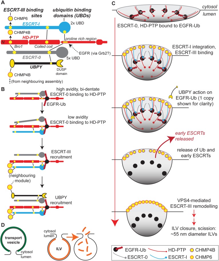
Figure 5. Identifying a potential mechanism for EGFR sorting to the MVB.
(A) Alignment shows the polarity of the MVB sorting machine. Key binding sites are shown in yellow. HD-PTP binding to EGFR may occur via Grb2, but other modes of interaction are possible. (B) Competitive binding reactions may explain how HD-PTP supports the sequential assembly of ESCRTs upon ubiquitinated EGFR. (C) A speculative model, in which these competitive binding reactions are superimposed onto the pathway of ILV formation. HD-PTP and ESCRT-0 attach to a core of ubiquitinated EGFR in a radial configuration. ESCRT-I integration helps expose ESCRT-III-binding sites towards the rim and may generate membrane curvature. From ESCRT-III, UBPY reaches cargo to destabilise the assembly. (D) Solving a topographical problem. Transport vesicle formation requires an exoskeleton (coat) (left). An ILV might use an ESCRT endoskeleton that is triggered to disassemble prior to membrane scission (right).

