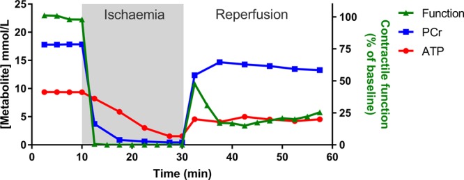
Figure 2. Ex vivo measurement of function and high-energy phosphates during no-flow I/R.

A classical Langendorff-perfused heart experiment (adapted and redrawn from ref. [49]). The heart is excised and perfused with an oxygenated physiological saline solution, whereupon it beats spontaneously. This can be performed in a magnet to simultaneously measure high-energy phosphates by 31P-NMR. After a period of equilibration, the perfusate is switched off to simulate ischaemia. Phosphocreatine (PCr) levels drop within seconds as they are preferentially used to maintain ATP, which only falls once PCr is exhausted. The effect on cardiac contractile function is observed within seconds and quickly falls to zero. ATP continues to reduce slowly during the ischaemic period, dependent on the balance between glycolysis and basal metabolic energy demands (e.g. ionic homeostasis), with accumulation of cellular calcium observed as an increase in end-diastolic pressure. Upon reperfusion, there is a very rapid recovery of PCr, which is critical to re-energising the cell and returning ionic homeostasis before long-term damage is induced. A post-reperfusion spike in function likely reflects high calcium levels, but it is the sustained functional recovery after 30 min reperfusion that is the main outcome measure.
