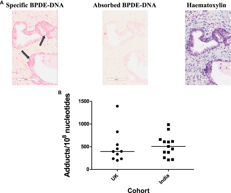
Figure 2. PAH-DNA immunostaining of human prostate.
(A) Representative example of PAH-DNA immunostaining in a UK prostate sample stained for carcinogenic PAH-DNA adducts: (left panel) specific PAH-DNA adduct staining is shown by nuclei stained pink and indicated by arrows; (middle panel) the corresponding absorbed serum control shows the same area with no staining and (right panel) haematoxylin staining of the same area shows localisation of nuclei. (B) Values for PAH-DNA adducts/108 nucleotides, for 10 prostate samples from the U.K. and 13 samples from India, were obtained from IHC using ACIS OD/nucleus values (with absorbed serum subtracted) by calculation from a standard curve [85,86].

