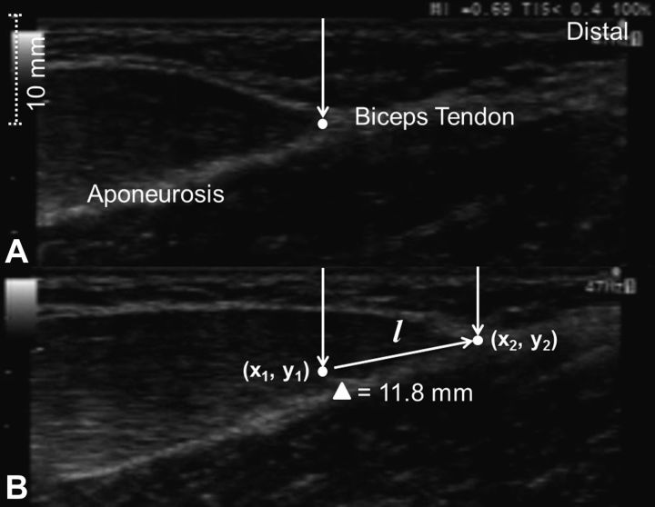
Fig. 1.
A typical B-mode ultrasound image of biceps brachii immediately before an eccentric contraction (maximal isometric contractions at 60° elbow flexion; A) and at the end of the eccentric contraction (at 0° elbow flexion; B). The displacement of the myotendinous junction (as shown by the white dot) was calculated from the two pictures based on the formula; , showing the myotendinous junction (as shown by an arrow) moved. The displacement (l) is 11.8 mm in this example.

