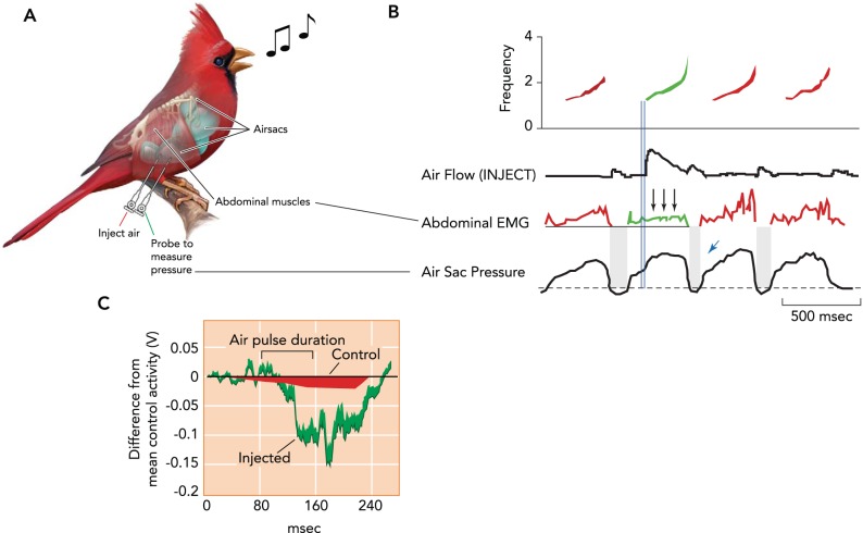
FIGURE 2.
Schematic of experimental approach and recorded parameters
Northern cardinals (Cardinalis cardinalis) show a compensatory response in expiratory muscles when a supplementary air pulse is injected into the air sac system during singing. A: schematic of the experimental approach and recorded parameters. Subsyringeal pressure (air sac pressure) is measured via cannulae (green line; cannula to measure pressure) implanted in the air sacs, and abdominal muscle (M. obliquus externus abdominus) activation is measured by electrodes placed in the muscle (Abominal EMG). Although the cardinal is singing, air is injected directly into the air sac through a second cannula (red line; Inject Air). B: when an air pulse is injected into a thoracic air sac at the beginning of a syllable (second of four syllables; shown in green), activation of the expiratory muscles is reduced during the remainder of the syllable (three arrows). Injection flow (onset illustrated by the double blue lines) is depicted in the trace below the spectrogram, with control syllables in red and the stimulated syllable in green. Abdominal muscle activation is shown as the rectified and integrated amplitude of the recorded EMG. The small blue arrow depicts the smaller than normal airflow and duration of the minibreath following injection of the air puff. C: quantification showing the reduction in relative mean amplitude of abdominal muscle activation when air was injected (green) compared with uninjected control syllables (red). The decrease in activation occurs with a delay (∼50 ms) after the onset of injection. Figure is modified from Ref. 66 with permission from Proceedings of the National Academy of Sciences USA.

