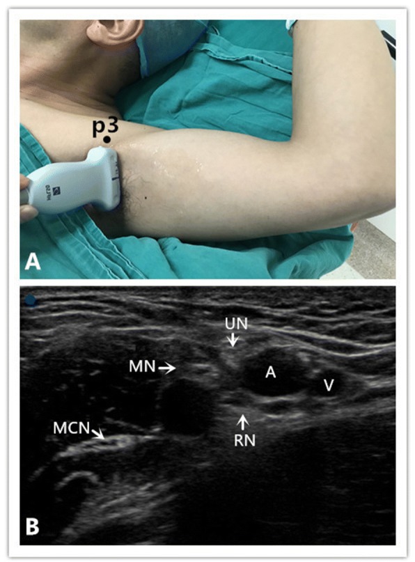
Fig. 2.

Position of patient and ultrasound imaging in the operating room when performing the axillary block. a Position of the patient, the probe, and the needle (“p3”) during the axillary block. b Ultrasound depiction of the axillary. Radial nerve (RN), musculocutaneous nerve (MCN), median nerve (MN), ulnar nerve (UN), axillary artery (A) and axillary vein (V) The blue point is situated at the lateral side of the probe
