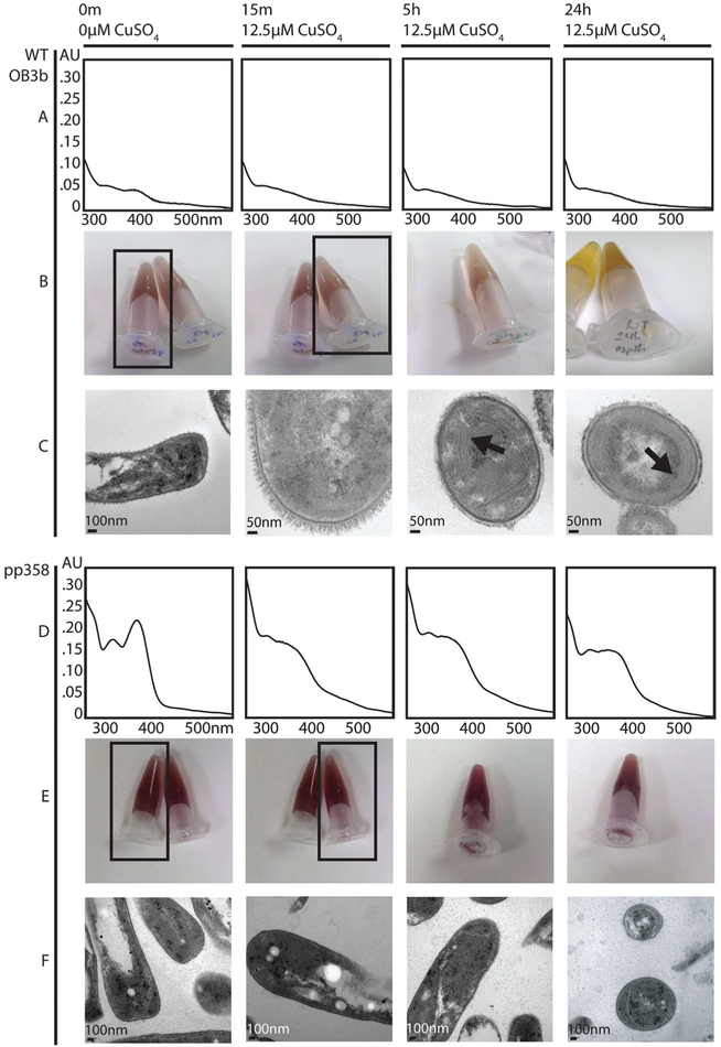
Fig. 3.
Phenotypic analysis of qRT-PCR samples. (A) Wildtype (WT) M. trichosporium OB3b spent medium UV-visible spectra. Apo Mbn is visible at 0 min (as shown by the peaks at 340 and 390 nm); addition of 12.5 μM CuSO4 to the medium results in chelation and a shift to a typical CuMbn spectrum. (B) Wildtype M. trichosporium OB3b naphthalene assay for sMMO activity. Oxidation of naphthalene by sMMO results in a pink/purple color in the presence of Fast Blue B; without sMMO activity, addition of Fast Blue B results in a primarily yellowish color. The relevant tubes for the 0 and 15 min timepoints are indicated by black boxes. (C) Transmission electron micrographs of wildtype M. trichosporium OB3b cells. By 5 h and 24 h, intracytoplasmic membranes are visible (indicated by arrows). (D) PP358 spent medium UV-visible spectra. Mbn is visible at 0 m and is more abundant than in wildtype M. trichosporium OB3b; addition of CuSO4 to the medium results in chelation and a shift to a typical CuMbn spectrum, but by 24 h features at 340 nm and 390 nm are reappearing, indicating that production of apo Mbn continues. (E) PP358 naphthalene assay for sMMO activity. Active sMMO remains present at all timepoints despite the addition of copper. (F) Transmission electron micrographs of PP358 cells. Significant numbers of intracytoplasmic membranes are not observed even at 5 h and 24 h.

