Figure 1.
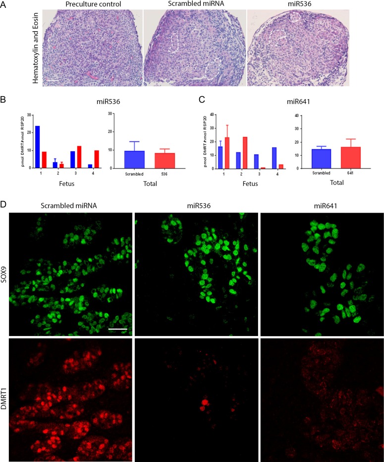
DMRT1 knockdown does not affect seminiferous cord formation in the first trimester human fetal testis.(A) H + E staining of pre-culture control (left panel) and after culture for 13 days following an initial 24 h lentiviral exposure to scrambled miRNA (middle panel) or miR536 (right panel). Seminiferous cords are delineated by broken white lines. DMRT1 mRNA expression in (B) miR536- and (C) miR641-exposed tissues compared to scrambled miRNA controls (n = 4 for each DMRT1-miRNA exposure group). (D) Immunofluorescence analysis of SOX9 (green) and DMRT1 (red) expression in Sertoli cells in fetal testis tissue following transduction with either scrambled miRNA (left panel), miR536 (middle panel) or miR641 (right panel) lentiviral constructs. Scale bars: (A) 20 μM and (D) 50 μM. Graphs show mean ± SEM. Data analyzed using a linear mixed effects model in R.

