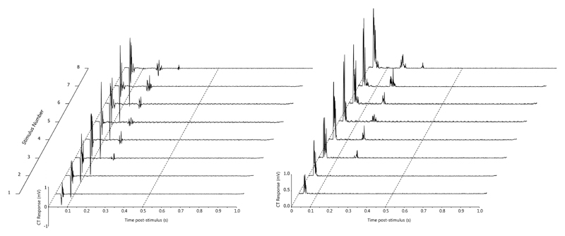
Figure 3.
An example of temporal summation in the cranial tibial muscle (recorded from dog 71). The top channel is a stimulus marker channel, with a train of 8 1ms 10 mA stimuli delivered at a frequency of 1 Hz. The lower channel shows the early and late responses in the cranial tibial muscle. The time base is 0.2s/division.

