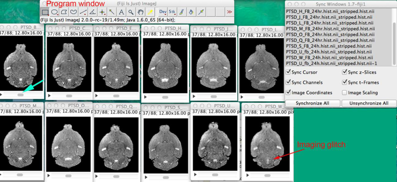
Figure 3. Visualization of MR images in FIJI/ImageJ 3D image viewer.

Open FIIJI and drag your set of MR images into the program window (top of screenshot above). Under the menu bar, pull down “Window” and select “Tile”. Then arrange the images on the screen. To synchronize all images so that you can scroll through the 3D stacks quickly, pull down the menu under “Analyze” select Tools>Synchronize windows. The Sync window will open (top right). You can now scroll any one image by moving the gray scroll button left or right under one of the images (turquoise arrow) and all will move synchronously through their respective stack. By watching the images, defects are apparent (red arrow).
