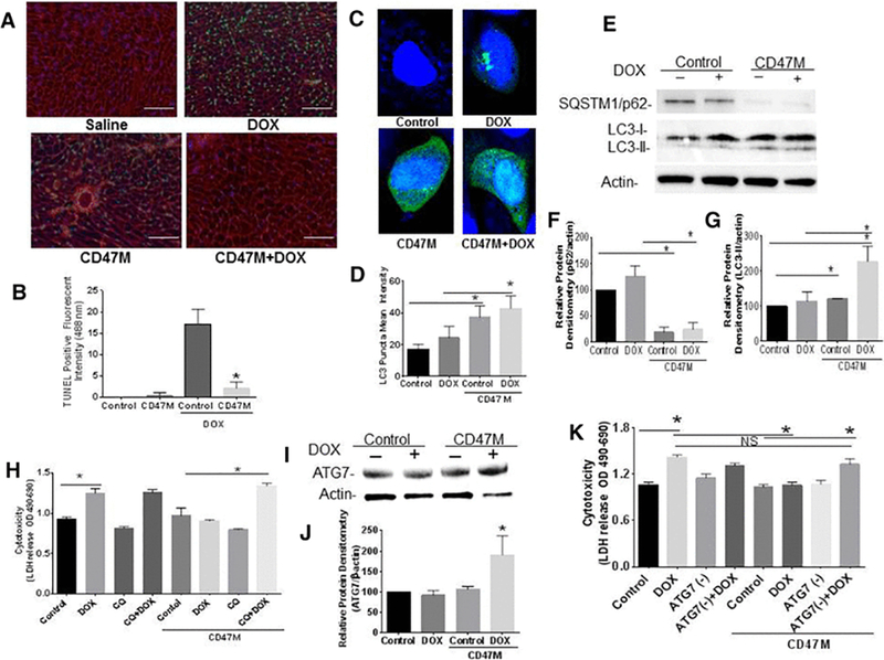
Figure 4. Blockade of CD47 protects cardiac myocytes and cardiac tissue from doxorubicin mediated toxicity.

(A) Hearts of mice were excised after doxorubicin treatment, sections were paraffin embedded and tissue was stained with TUNEL and WGA (red) to detect apoptotic cardiac cells (40x, Scale bar= 100μm). Autophagic flux was measured by expression of SQSTM1/p62 and LC3 lipidation by Western Blot hybridization (B) and quantified (C, D n=4 *p<0.05). H9C2 cardiac myoblasts were treated with CD47M and or DOX and transfected with LC3-GFP LC3 puncta was determined by confocal microscopy (E) and quantified using ZEN 2.3 SP1 software (F). H9C2 cardiac myoblast cells were plated and left untreated or administered CD47 Morpholino (CD47 M); 48 h after cells were treated with or without DOX in the presence or absence of 25 μM chloroquine cells were incubated 48 h and LDH release was measured to determine cell cytotoxicity n=4 *p<0.001 (G).
