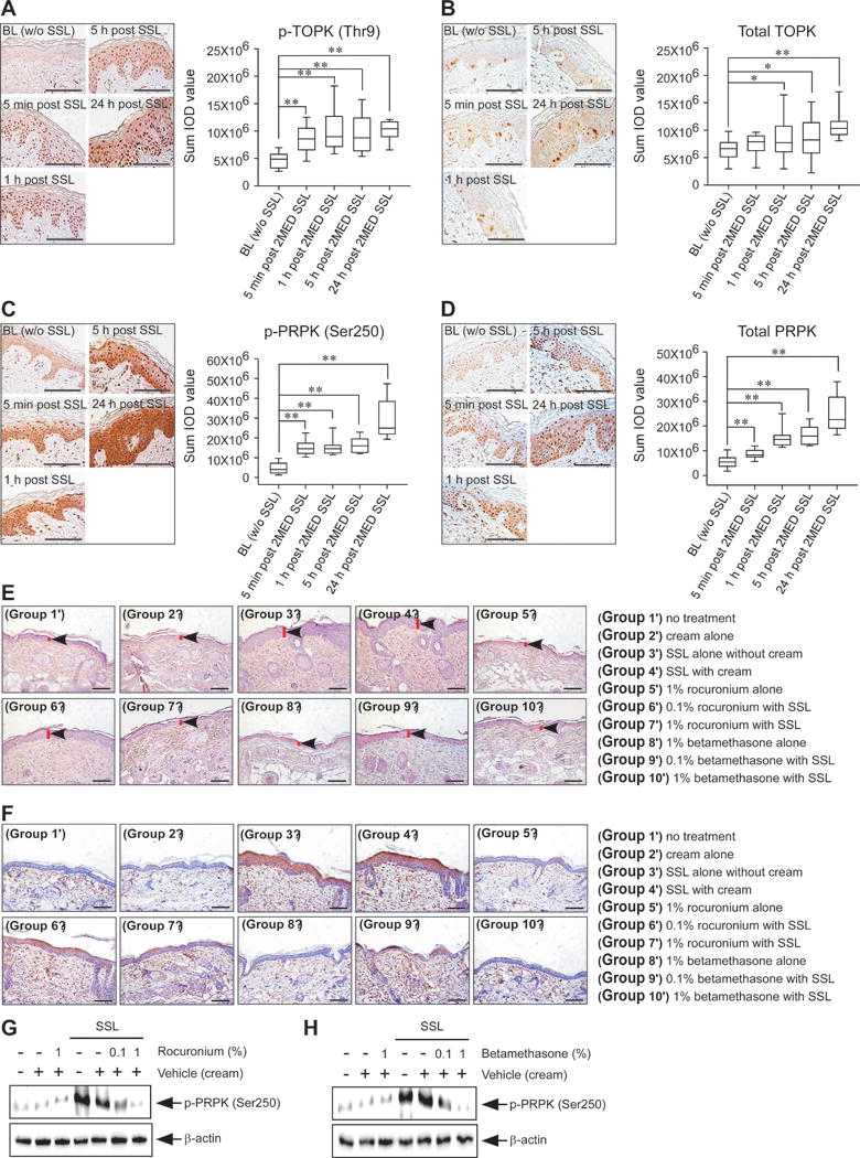
Figure 4. Effects of PRPK inhibitors on acute SSL-induced inflammation in human or mouse skin.

For clinical human tissues, the minimal erythemic dose (MED) was defined as the smallest dose of energy necessary to produce confluent erythema with four distinct borders at 24 h post-exposure. MED was determined on a buttock area previously unexposed to sunlight. Each test area was subdivided into six sub-sites (each 1 cm2) corresponding to the liquid light guide pattern on the solar simulator. The solar simulator was calibrated prior to each use and a series of six increasing SSL irradiation exposures were administered to each sub-site area. Following exposure, the test sites were covered until evaluations were completed. After determination of the MED for each individual, the contralateral buttock was exposed to twice the determined MED. A 6-mm skin punch biopsy sample was collected from one buttock at baseline prior to SSL exposure and additional 4 mm punch biopsies were removed at 5 min, 1, 5, and 24 hours post-SSL irradiation. IHC analysis of normal human skin (BL) without SSL exposure (30 photos from 10 human subjects), 5 min post 2 MED SSL exposure (30 photos from 10 human subjects), 1 h post 2 MED SSL exposure (30 photos from 10 human subjects), 5 h post 2 MED SSL exposure (30 photos from human 10 subjects) and 24 h post 2 MED SSL exposure (30 photos from 10 human subjects) to detect expression of (A) phosphorylated TOPK (Thr9), (B) total TOPK, (C) phosphorylated PRPK (Ser250) or (D) total PRPK. Phosphorylated TOPK, total TOPK, phosphorylated PRPK and total PRPK levels are presented as sum of IOD values (right panel in each figure). Left panel in each figure shows representative staining (brown color). The asterisks (*, **) indicate a significant (p < 0.05 or p < 0.01, respectively) difference compared to the normal human skin (BL). (E) At 24 h after SSL irradiation, mouse skin tissues were prepared for H&E staining to compare the epidermal thickness. (F) IHC and (G, H) Western blot analyses were conducted to examine the effect of rocuronium bromide and betamethasone 17-valerate on phosphorylated PRPK levels in acute SSL-stimulated SKH1 hairless mouse skin. Groups were divided for Figure 4E–H as below; Group 1′ = no treatment; Group 2′ = cream alone; Group 3′ = SSL alone without cream; Group 4′ = SSL with cream; Group 5′ = 1% rocuronium bromide alone; Group 6′ = 0.1% rocuronium bromide alone; Group 7′ = 1% rocuronium bromide with SSL; Group 8′ = 1% betamethasone 17-valerate alone; Group 9′ = 0.1% betamethasone 17-valerate with SSL; and Group 10′ = 1% betamethasone 17-valerate with SSL. Scale bars are 100 μm for Figure 4A–F.
