Figure 1.
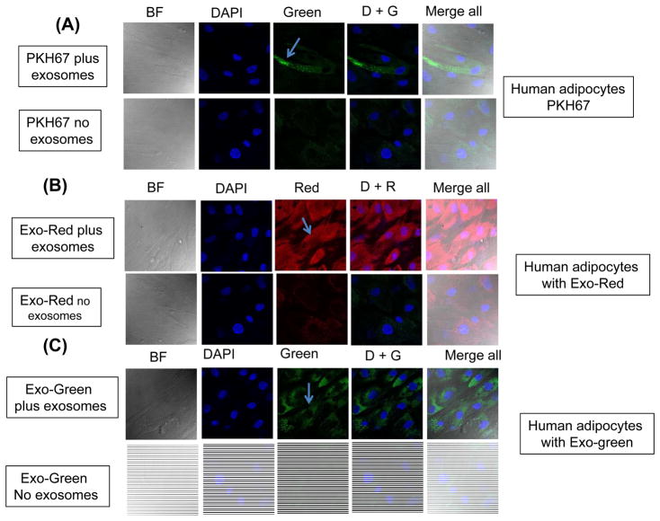
Uptake of fluorescently labelled exosomes by differentiated human adipocytes cells. Confocal microscope images illustrating exosome uptake into human adipocytes cells. Exosomes were isolated from plasma of subjects with obesity hypoventilation and labeled with the PKH67 Green Fluorescent (lipophilic), Exo-Red (fluorescently-label isolated exosome RNAs), and Exo-Green (fluorescently-label isolated exosome protein). Panel (A) is a representative images of human differentiated adipocytes cells were grown on coverslips for 24 h and the labeled exosomes with PKH67 were added to the cells for 6 h at 37°C. Panel (B) a representative images of human differentiated adipocytes and the labeled exosomes with Exo-Red were added to the cells for 6 h at 37°C. Panel (C) is a representative images of human differentiated adipocytes and the labeled exosomes with Exo-Green were added to the cells for 6 h at 37°C. Cells were washed and stained with nuclei (blue) stained with DAPI, n=6, scale bar in 10 μm. As controls, no exosomes were used but PKH67 was added. The differential interference contrast (DIC) was used to visual the morphology of the cells without fluorescence.

