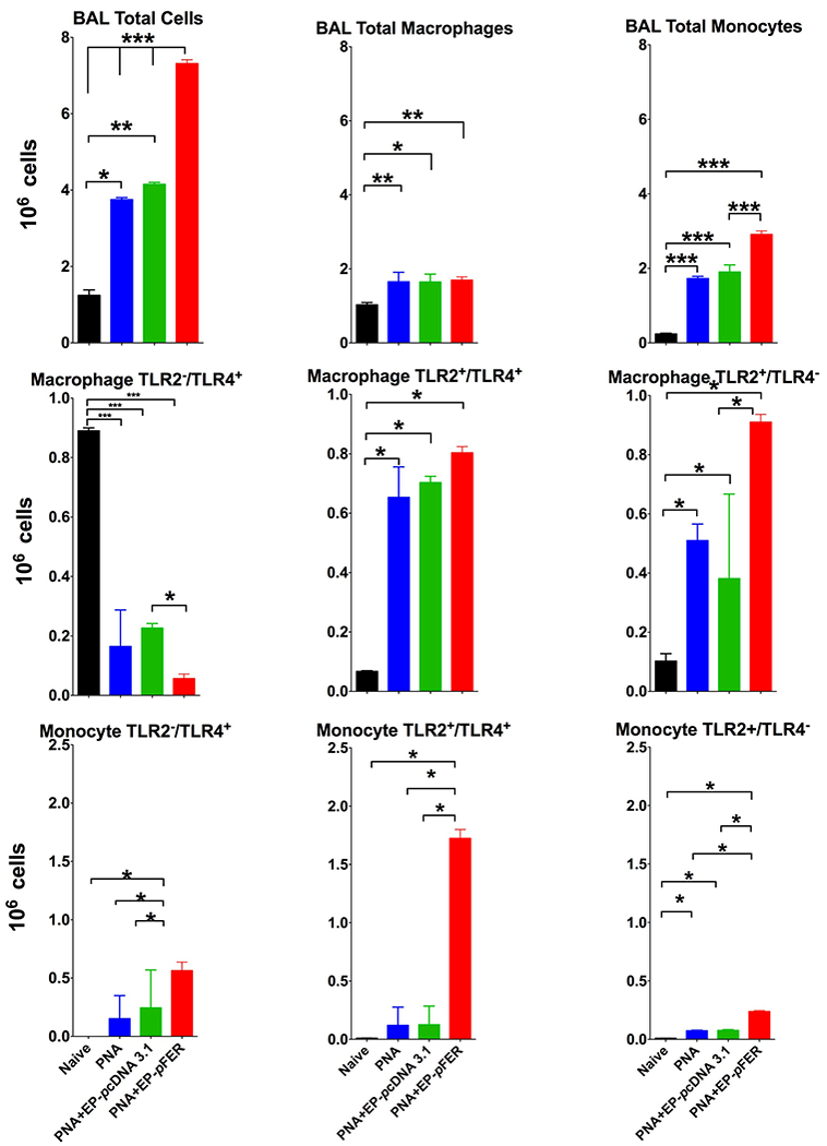
Figure 4. Flow cytometry analysis of bronchial alveolar lavage fluid (BAL) shows robust recruitment of inflammatory monocytes and modest recruitment of macrophages after FER electroporation treatment.

BAL fluid was recovered and cells were stained for Gr-1, F4/80, CD11b and CD206. Top Row Total number of BAL cells were higher in the FER treated group compared to controls (PNA-only, PNA EP-saline) being the inflammatory monocytes the main driver in this difference. Middle Row. Cell sorting of macrophages showing with concomitant TLR-2 and TLR-4 staining incremented by FER treatment showed increased proportion of TLR2+/TLR4+ and TLR2+/TLR4− cells. Bottom Row Cell sorting of inflammatory monocytes with further staining for Toll-like receptors 2 and 4 showed a very significant increase of TLR2+/TLR4+ as compared to other phenotypes. N=10. One-way ANOVA with Tukey’s multiple comparison tests (* p < 0.05; ** p < 0.01; *** p < 0.001).
