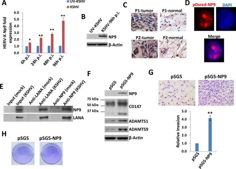
Figure 4. KSHV infection induces HERV-K encoded oncogenic NP9 expression which enhancing primary endothelial cells invasion and colony formation.

(A-B) HUVEC were infected with purified KSHV (MOI~10) or UV-inactivated KSHV for 2h, then the induction of HERV-K NP9 at indicated time-points post-infection (p.i.) was measured and compared to UV-inactivated KSHV infected cells control using qRT-PCR and immunoblots. (C) The strong expression of NP9 protein in KS tissues from our cohort of two AIDS-KS patients without any treatment by immunohistochemistry staining. (D-E) HUVEC were transfected with the pDsred-NP9 vector for 48 h, then protein expression was detected by immunofluorescence and nuclear was shown by DAPI. Immunoprecipitation assays in both directions were performed using Catch and Release Immunoprecipitation Kit (Millipore) with anti-LANA or anti-NP9 antibodies, respectively. (F-H) HUVEC were transfected with pSG5 control vector or pSG5-NP9 for 48 h, then protein expression was detected by immunoblots. Cell invasiveness was determined using the transwell assays. Anchorage-independent growth ability was determined using the soft agar assays. Error bars represent the S.D. from 3 independent experiments. * = p<0.05, ** = p<0.01.
