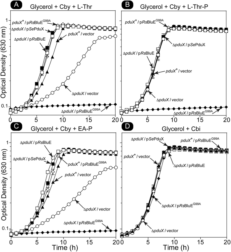
Figure 5. RsBluEG99A variant disrupts all the enzymes in the entire AP-P synthesis and attachment branch in S. enterica in vivo.
Representative graphs of growth analyses of S. enterica cells grown aerobically at 37°C in NCE minimal medium with glycerol (22 mM) ampicillin (0.1 mg mL−1), and MgSO4 (1 mM) Cby (1 nM) and supplemented with A. L-Thr (1 mM), B. L-Thr-P (1 mM), or C. ethanolamine phosphate (EA-P, 1 mM). Cbi (1 mM) was used as the corrinoid in place of Cby in panel D. Experiments were replicated in two independent experiments, each performed in triplicate. Error bar represent the standard error of the mean. Figure key: ΔpduX/pRsBluE (□), ΔpduX/pSePduX (■), ΔpduX/pRsBluEG99A (◆), pduX+/pRsBluEG99A (◇), pduX+/vector (▲), ΔpduX/vector (○).

