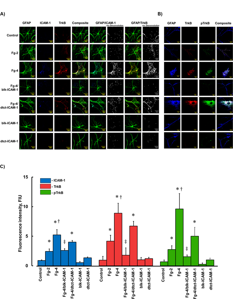
Figure 2. Fibrinogen (Fg) induced overexpression of intercellular adhesion molecule-1 (ICAM-1), tyrosine receptor kinase B (TrkB) and its phosphorylation (pTrkB).

A and B) Examples of images of cultured mouse brain astrocytes (MCAs) treated with serum free media alone (control), 2 mg/ml of Fg (Fg-2), 4 mg/ml of Fg (Fg-4), 4 mg/ml of Fg in the presence of function blocking anti-ICAM-1 antibody (Fg-4/blk-ICAM-1), 4 mg/ml of Fg in the presence of detecting anti-ICAM-1 antibody (Fg-4/dtct-ICAM-1), or with function blocking or detecting anti-ICAM-1 antibodies alone (blk-ICAM- 1 and dtct-ICAM-1, respectively).
A) Astrocytes were identified by expression of glial fibrillary acidic protein (GFAP, green); ICAM-1 -blue, and TrkB -red.
B) Astrocytes were identified by GFAP -blue, TrkB -red, and pTrkB - green.
C) Summary of fluorescence intensity changes for ICAM-1, TrkB, and pTrkB in cultured MCAs treated with Fg-2, Fg-4, Fg-4/blk-ICAM-1, Fg-4/dtct-ICAM-1, blk-ICAM-1, or dtct-ICAM-1.
P < 0.05 for all. * - vs. Control, †- vs. Fg-2, ‡- vs. Fg-4; n=4
