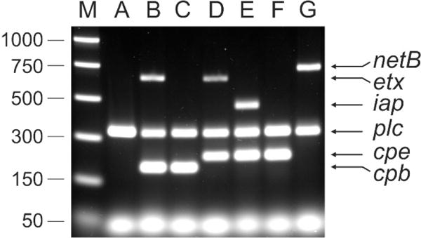
Fig. 1. Multiplex PCR analysis of representative C. perfringens type A to G strains.

The strains were grown in TGY broth [79] to a turbidity at 600nm between 1.0 and 1.5. Genomic DNA (equivalent to 5mL of culture) was prepared as described previously [80]. DNA preparations were diluted 1 in 50 in sterile distilled water and used as templates in the multiplex toxin PCR based on a previous method [81]. The oligonucleotide primers and their concentrations are listed in Table 2. PCR reactions were prepared using 0.1 units/μL Taq DNA polymerase (Roche) in 1 x supplied buffer (Roche), 2 mM MgSO4 and 0.4 mM dNTPs. The template constituted 0.1 volumes of the final reaction. PCR was performed with an initial denaturation at 95°C for five minutes, followed by 35 cycles of 95°C for 1 minute, 55°C for 1 minute and 72°C for 1 minute. Amplified products were resolved by electrophoresis through a 1.5% (w/v) TAE agarose gel. The multiplex PCR profiles of the following C. perfringens strains are shown: JIR325 (type A)[82], JGS1984 (type B)[83], CN3717 (type C)[84], JGS4138 (type D)[85], ATCC27324 (type E), SM101 (type F)[86], EHE-NE18 (type G)[87]. Size standards were PCR Markers (Promega).
