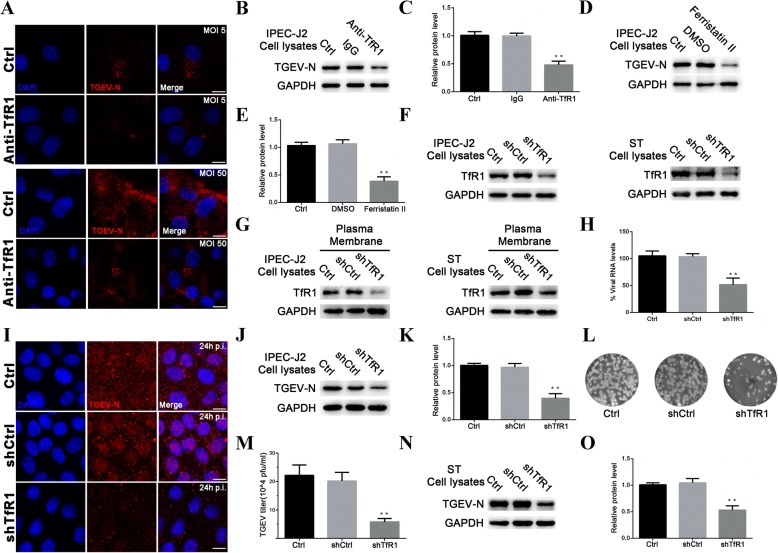
Fig. 3.
Blocking and Suppressing TfR1 protein inhibits TGEV Infection. a IPEC-J2 cells were pre-incubated with anti-TfR1 Ab for 1 h at 37 °C. Early invasion of TGEV (at 1 h p.i.; MOI 5 or 50) was inhibited. b and c IPEC-J2 cells were pre-incubated with anti-TfR1 Ab for 1 h at 37 °C. Replication of TGEV-N protein was detected at 24 h p.i. (MOI 5) by western blotting. d and e IPEC-J2 cells were pretreated with 50 μM ferristatin II and then infected with TGEV (MOI 5). For controls, cells that had not been pretreated with ferristatin II were infected with TGEV or pretreated with DMSO. Cell lysates were harvested for western blotting at 24 h. f and g Interference with total and membrane TfR1 levels in IPEC-J2 and ST cell lines were verified by western blotting. h IPEC-J2 cells were transfected with interference vector pLVX-shRNA-TfR1. pLVX-shRNA-Ctrl and normal cells served as controls. 1 h after infection with TGEV (MOI 5), TGEV-N mRNA levels were quantified by RT-PCR. i Confocal images of TGEV (MOI 5) infected IPEC-J2 cells transfected with shTfR1. Images were obtained 24 h p.i. The red stain indicates TGEV-N. Nuclei are stained with DAPI (blue). Scale bar = 10 μm. j and k shRNA was used target TfR1 in IPEC-J2 cells. The cells were infected with TGEV (MOI 5), and analyzed at 24 h p.i. by western blotting. l and m Culture supernatants from Fig j were harvested and virus titers were measured by plaque assay in ST cells. Plaques developed at 2 days after infection. Untreated cells and cells transfected with scrambled shRNA cells served as controls. n and o ST cells stably carrying the shTfR1 interference vector were infected with TGEV (MOI 5) and harvested at 24 h p.i. for western blotting. TGEV replication was inhibited by TfR1 knockdown. The TGEV-N to GAPDH ratio was normalized to control conditions. The data shown are means ± SD from three independent experiments. (* 0.01 < p < 0.05, ** p < 0.01)

