Fig. 4.

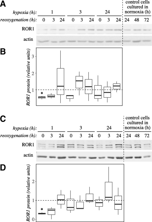
ROR1 protein level is regulated in cardiomyocytes in response to hypoxia and reoxygenation. a A representative Western analysis of ROR1 protein level in HL-1 cardiomyocytes after treatment with hypoxia and reoxygenation. Cells were allowed to adhere for 24 h after plating in normoxia. This was followed by culturing the cells in a hypoxic work station at 1% O2 (hypoxia) and subsequently again in the regular cell incubator in normoxia (reoxygenation) for the indicated periods of time. As time points were distributed over three days after plating, control samples cultured in normoxia for 24, 48 or 72 h were also analyzed. Time points (hypoxia+reoxygenation) 1 + 0, 1 + 3, 3 + 0 and 3 + 3 are comparable to the 24 h control (lane 10), time points 1 + 24, 3 + 24, 24 + 0 and 24 + 3 to the 48 h control (lane 11), and time points 24 + 24 to the 72 h control (lane 12). b A box plot presentation of quantitation of ROR1 bands from three Western blots similar to the one shown in panel A. ROR1 band intensities were normalized to each sample’s actin level, and subsequently divided by the control sample value of the respective timepoint. c A representative Western analysis of ROR1 protein levels in H9c2 cardiomyoblasts after treatment with hypoxia and reoxygenation. Experiment was carried out as shown for HL-1 cells in panel A. The antibody recognized two bands between 130 and 180 kDa, that both were down-regulated by ROR1 siRNA knockdown (data not shown). d A box plot presentation of quantitation of ROR1 bands from three Western blots similar to the one shown in panel C. Data were normalized as for panel B. Asterisk indicates significant difference in expression (P < 0.05) as compared to control samples
