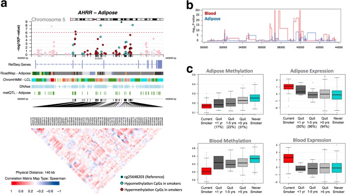
Fig. 5.
Tissue-shared smoking-associated DNA methylation and gene expression patterns at AHRR. a coMET plot [49] of the association between 66 AHRR CpG sites and smoking. Top panel shows the -log10P value of the association; the middle panel shows genomic annotation; and the lower panel shows co-methylation patterns based on Spearman correlation coefficients. b Tissue-shared and tissue-specific methylation signals across CpG sites in the AHRR gene region in adipose (blue) and blood samples (red). c DNA methylation and gene expression levels with respect to smoking cessation. Methylation and gene expression levels are shown for five different smoking status categories: current smokers (red); subjects who quit within 1 year, subjects who quit between 1 and 5 years, and subjects who quit over 5 years at the time of methylation sampling (gray); and non-smokers (blue). X-axis labels include the proportion of subjects who reverted in each smoking quit year category

