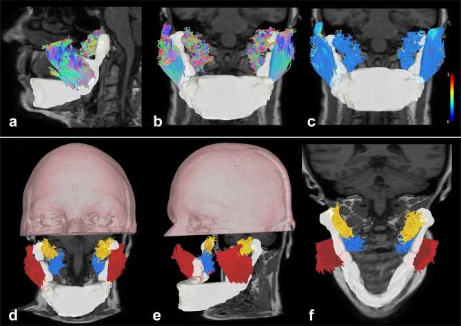
Figure 1.
Tractographic reconstructions of masticatory muscular complex. (a, b) Sagittal and frontal view of the masticatory muscular complex with a directional color coding representation, overimposed on a T1 weighted image (the mandible is reconstructed as a white volume). (c) Frontal view of the masticatory muscular complex with a local index of FA color coding representation overimposed on a T1 weighted image (lateral scale represents FA values from 0 to 1). (d–f) Volume rendering of the upper portion of the head of the patient with tractographic reconstructions of masticatory muscular complex (axial and coronal T1 weighted images are underlaid). Arbitrary colors were given to fibers: masseter muscles are represented in red, lateral pterygoid muscles are represented in yellow and medial pterygoid muscles are represented in blue.

