
Figure 2.
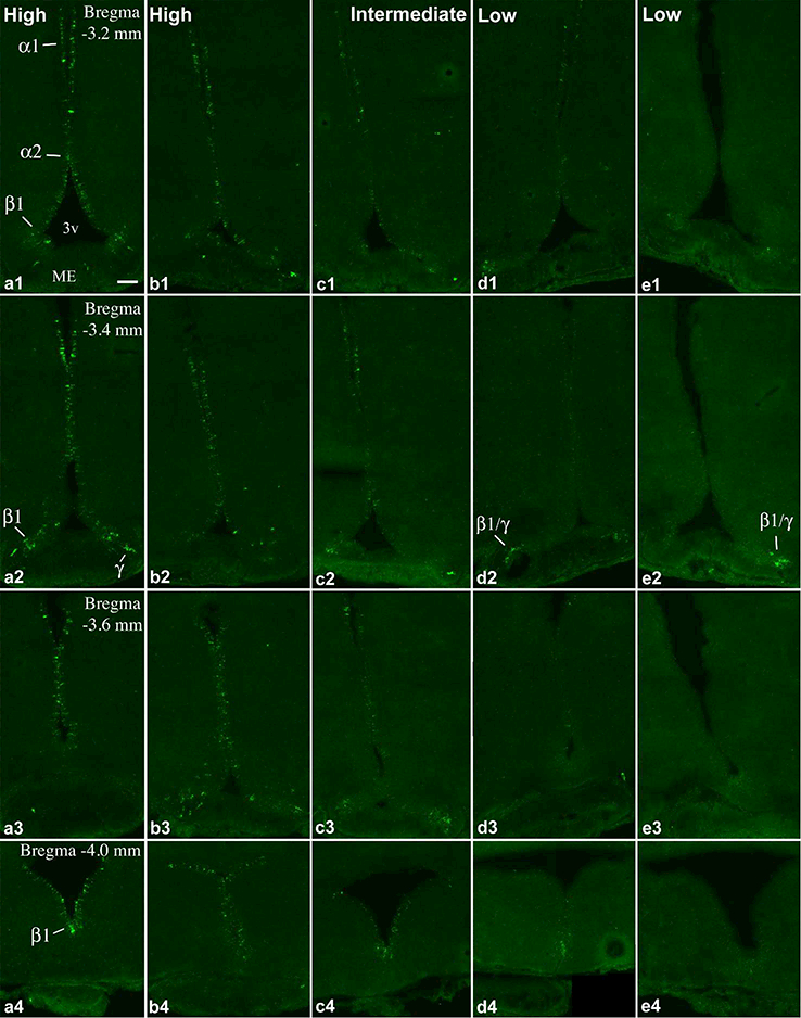
Continuation of Figure 1. FISH demonstrates Prss56 mRNA-expressing cells in the caudal part of the tanycyte region in 5 adult rats with different expression levels. (a, b) High, (c) intermediate, (d, e) and low Prss56 mRNA levels in tanycytes. Pomc expression in brains b, d and e, are shown in A, D and E, respectively, of Figs. 1 and 2 in Wittmann et al. (2017). Tanycyte subtypes (α1, α2, β1 and γ) that express Prss56 are indicated on a1, a2, a4, d2, e2. 3v, third ventricle; ME, median eminence. Scale bar: 100μm.