
Figure 9.
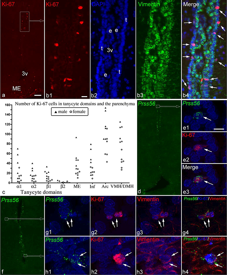
(a) Ki-67 immunofluorescence demonstrates a highly proliferative zone in the α1 tanycyte domain (boxed area). (b1–4) Higher magnification images of this zone show that most Ki-67 cells are in the layer of tanycytes (t in B2, nuclear DAPI staining) facing the parenchyma, and express vimentin (green; arrows in b4). 3v, third ventricle; e, ependymal cell layers. (c) The number of Ki-67 cells shows large variations among 6 male and 6 female rats in tanycytic domains, the median eminence (ME), infundibular stalk (Inf), arcuate nucleus (Arc), and the ventromedial and dorsomedial nuclei (VMH/DMH). (d-h) Ki-67 in Prss56-expressing cells (white arrows), including an α1 tanycyte (d, e), parenchymal cells (f, g) and β1 tanycytes (f, h). Vimentin in the cytoplasm of the parenchymal cells and β1 tanycytes is shown in g3 and h3. Scale bar: 100μm in a (for a, d, f); 20μm in b; and 10μm in e1 (for e, g, h).