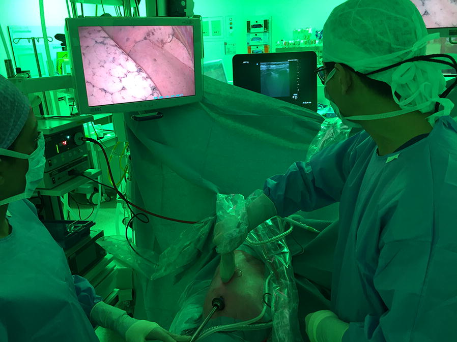
Fig. 2.

The surgeon performs lung ultrasonography in a patient with a confirmed pneumothorax and the videoscope in situ. The video screen displays an image of the inside of the right hemi-thorax and the collapsed right lung. The surgeon is handling the wrapped-up ultrasound transducer. The ultrasound device is shown in the back of the image
