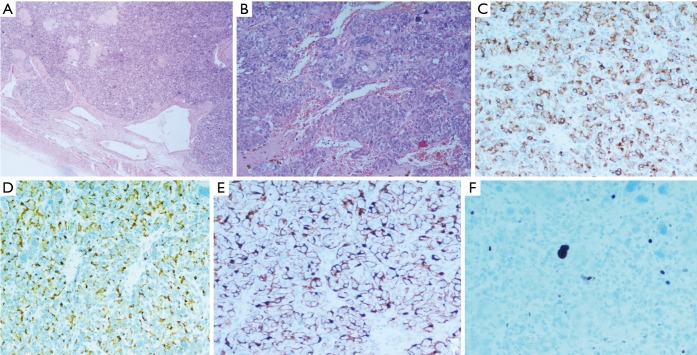
Figure 4.
Mediastinal paraganglioma: (A) haematoxylin-eosin 100× showing the peripheral capsule and a rich microvasculature; (B) haematoxylin-eosin 400× showing a typical pattern of nests of neoplastic epithelioid cells, with large hyperchromic nuclei, surrounded by thin-walled dilated vessels; (C) positive immunohistochemical staining for chromogranin A in the neoplastic cells, mostly prevalent at the perinuclear site (200×); (D) positive immunohistochemical staining for synaptophysin in almost 90% of the neoplastic cells, mostly prevalent at the cellular membrane (200×); (E) positive immunohistochemical staining for S-100 protein in the cytoplasm of the sustentacular cells, drawing a delicate net around the neoplastic cells (200×); (F) few neoplastic cells stain positively for Ki-67 (200×); note that many large hyperchromic nuclei do not result positive, revealing the degenerative nature of the phenomenon.

