Figure 5.
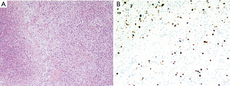
Pulmonary paraganglioma: (A) haematoxylin-eosin 100× showing a typical pattern of nests of monomorphic neoplastic cells, with scarce cytoplasm; an area of neoplastic coagulative necrosis is visible on the left; (B) a high number of proliferating neoplastic cells stain positively for Ki-67 (200×) and in this patient was associated with a negative prognosis.

