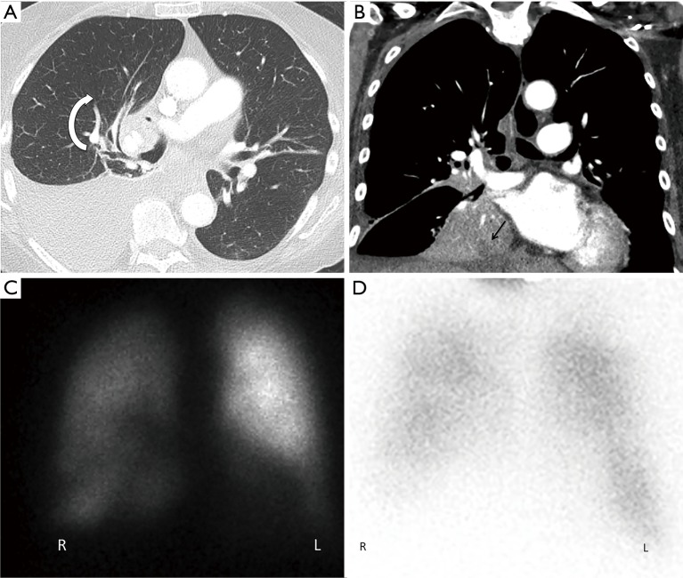
Figure 1.
Thoracic Imaging of pulmonary torsion. (A) Axial chest CT revealed clockwise swirling around the right hilum with distortion of the bronchovascular bundle. Note moderate right pleural effusion. (B) Coronal images revealed inferomedial rotation of the collapsed right upper lobe with internal hypoattenuation representing a juxtadiaphragmatic abscess (arrow). (C) Anterior Tc-MAA perfusion image demonstrated markedly decreased right lung perfusion compared to the left. (D) Corresponding Xenon-133 ventilation scan showed right lung hypoventilation.

