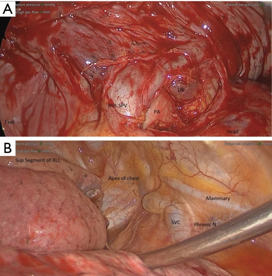
Figure 3.

Intraoperative anatomy. (A) Intraoperative anatomy after dissecting the pleura from hilum and untwisting the RUL. The RUL SPV is caudally displaced, while the azygous vein is displaced cranially. (B) The entire lung is rotated 180 degrees clockwise, as demonstrated by the RLL resting in the apex of the chest. LN, lymph node, RUL SPV, right upper lobe superior pulmonary vein; PA, pulmonary artery; SVC, superior vena cava; phrenic N, nerve; RLL, right lower lobe.
