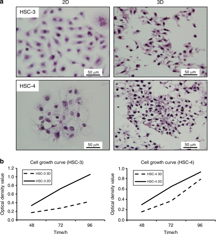
Fig. 2.
Differences in the morphology of 2D- and 3D-cultured HSC-3 and HSC-4 cells. a Cells cultured for 3 days on glass coverslips were mostly rounded. In contrast, protrusions were noted in the margin of cells 3D-cultured on Cellbed. HE staining (×400; scale bar: 50 μm). b MTT solution was added to the medium after incubation for 48, 72 or 96 h, and absorption was measured at each time point. We confirmed cell survival by MTT assay in both 2D and 3D culture conditions

