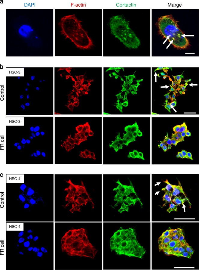
Fig. 7.
Immunofluorescent staining of 2D and 3D-cultured tongue cell lines. a Immunofluorescent staining was carried out on HSC-3 cells after 2D culture for 2 days on a glass coverslip. The colocalization of F-actin and cortactin, known as invadopodia, was observed in the form of punctae in the cell cytoplasm (scale bar: 10 μm). b, c Immunofluorescent staining of 3D-cultured tongue cell lines. Cell projections formed in the control cells in both HSC-3 (b) and HSC-4 cell lines (c); F-actin and cortactin colocalized in the cell margin. The formation of cell projections was markedly decreased in FR cells (scale bar: 50 μm)

