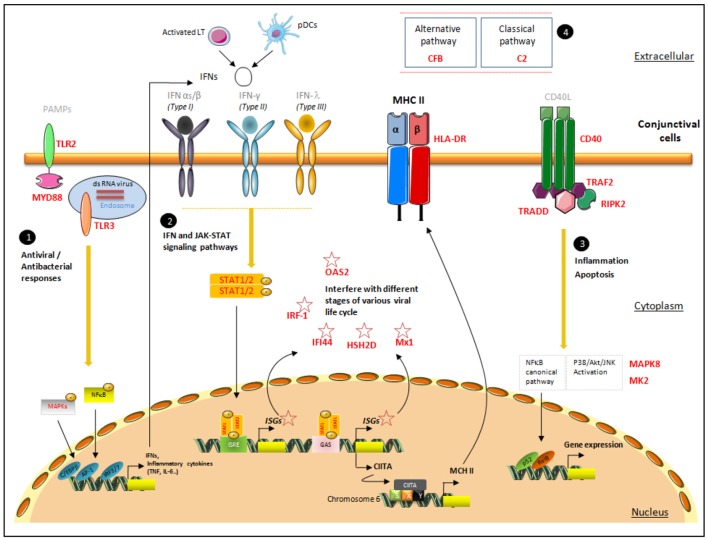
Figure 3.
Proposal for a pattern of signaling pathways associated with increased HLA-DR expression in conjunctival cells. This figure presents the localization of the targets identified with their possible interactions in the conjunctiva's spatial microenvironment. The four major signaling responses, in black circles, are mediated by (1) TLR responses, (2) IFN responses, (3) TNF responses, and (4) members of complement pathways. The target genes selected via their high correlation with HLA-DR are mentioned in bold red. The first line of pathogen recognition could be mediated by TLR responses via MAPK and NF-κB to induce inflammatory cytokine responses as INFs. IFNs bind to their receptors and initiate a signaling cascade, involving the JAK-STAT family of transcription factors, which leads to the transcriptional induction of the ISGs (IRF-1, IFI-44, HSH2D, Mx1, and OAS2), class II transactivator (CII-TA), and HLA-DR complex, which will migrate to the membrane. CD40, TRAF2, TRADD, and RIPK2, involved in TNF pathways, promote NF-κB and the MAPK family: MK2 and MAPK8, members of the p38 MAPK and JNK cascades, respectively. Pathogen-associated molecular patterns (PAMPs); CCAAT/enhancer binding protein beta(C/EBPβ); AP-1 transcription factor subunit(AP−1); IFN regulatory factor IRF-3 and IRF-7(IRF3/7); IFN-stimulated response element (ISRE); IFNγ-activated site (GAS); phosphate(P); class II transactivator (CIITA); MHC class II-specific regulatory module (XYS); nuclear factor kappa B subunit 2(p52); NF-κB subunit transcription factor RelB (RelB).

