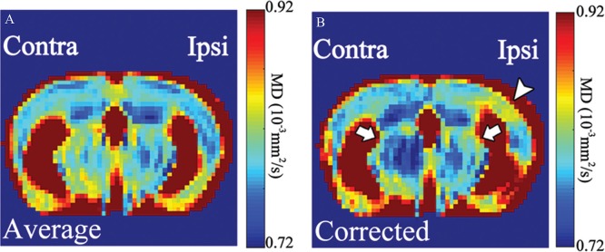
Fig. 4.
Maps of the average mean diffusivity (MD) before (A) and after (B) correction for the influence of blood flow. Mean diffusivity on the ipsilateral side appears higher than the contralateral side in dorsal cortex and corpus callosum with external capsule (CC + EC) (B: arrowhead). It may also be higher in the hippocampus (B: arrows).

