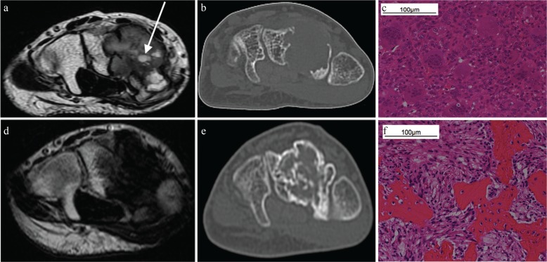
Fig. 2.
Computed tomography and MRI of a 33 y/o male with giant cell tumors of the bone (GCTB) in the carpal bones. Images are shown before and after denosumab treatment. (a and b) The tumor exhibited a well-defined geographic lucent lesion in the carpal bones that was 25 mm in size. On T2-weighted axial imaging, the tumor demonstrated well-circumscribed inhomogeneous hypo-intensity and a small cystic component (arrow). Computed tomography did not show bone formation in the tumor. (c) Both multinucleated giant cells and intervening mononuclear cells were observed in this photomicrograph. (d and e) On T2-weighted axial imaging, the tumor exhibited inhomogeneous hypo-intensity with an unclear boundary. The small cystic component disappeared after treatment. The tumor had slightly decreased in size to 22 mm. Computed tomography revealed GCTB in the carpal bones and new bone formation within the tumor. (f) Intermixed bone and fibroblast-like spindle cells were observed instead of multinucleated giant cells and intervening mononuclear cells.

