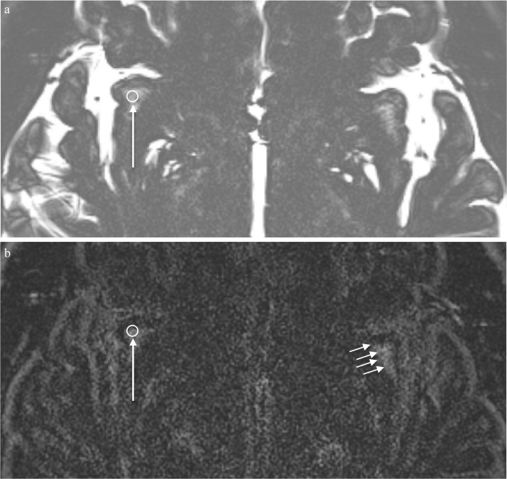
Fig. 1.
A 31-year-old healthy man. An example of the ROI placement in the perivascular space of the right subinsular white matter. (a) A 3 mm circular ROI is placed to include the structures with high signal intensity in the subinsular white matter on the axial MR cisternography (MRC) image parallel to the anterior commissureposterior commissure (AC-PC) line (arrow). This ROI is copied onto the corresponding heavily T2-weighted 3D-fluid attenuated inversion recovery (FLAIR) image (b) to measure the signal intensity (long arrow). Similarly, an ROI is placed on the other side and both sides were averaged for use in further analysis. (b) A corresponding heavily T2-weighted 3D-FLAIR image obtained prior to contrast administration. The ROI was copied from the MRC image (long arrow) to the subinsular white matter of the right side. Multiple linear or featherlike structures representing the perivascular spaces in the white matter in the corresponding left side subinsular area show high signal intensity even on this non-contrast enhanced image (short arrows).

