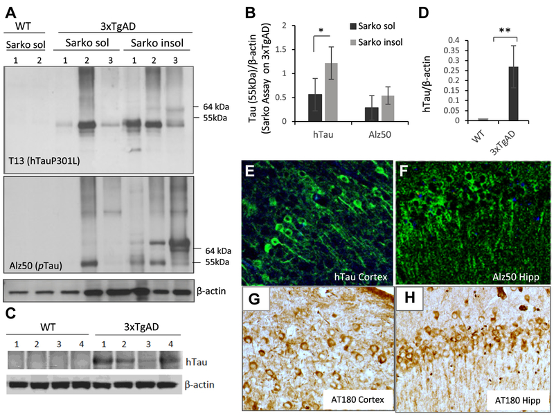
Fig. 4.
Characterization of tauopathy in 3 × TgAD mice. (A) Immunoblots of cerebral cortex tissue samples from 1-year-old WT and 3 × TgAD mice. Blots of samples from the sarkosyl-soluble or -insoluble fractions were probed with antibodies against mutant human tau (upper blot; 55 kDa band), phospho-tau (Alz50 antibody) (middle blot), and β-actin (lower blot). The band at 55 kDa corresponding to human tau was quantificated. (B) Results of quantification of sarkosyl-soluble and -insoluble hTau in cerebral cortex tissue samples from 1-year-old 3 × TgAD mice. *p < 0.05. (C) hTau protein immunoreactivity is not detectable in cerebral cortex tissue from WT mice by standard Western blot with anti-hTau antibody. (D) Results of quantification of mutant hTau levels in cerebral cortex tissues from WT and 3 × TgAD mice. **p < 0.001. (E–H) Images showing immunoreactivity of neurons with the human tau antibody and phospho-tau antibody Alz50 and AT180 in the Hip and cerebral cortex of a 1-year-old 3 × TgAD mouse. Abbreviations: AD, Alzheimer’s disease; Hip, hippocampus; WT, wild type.

