
Figure 1.
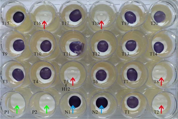
Screening of QSI strains on a 24‐well plate. The plate contained the biosensor strain, C. violaceum 12472 and filter paper with an aperture of 0.22 μm was used for sample detection. LB medium (blue arrows) and furanone (green arrows) served as negative and positive controls respectively. The absence of purple, or the formation of pigment inhibition, was considered indicative of potential QSI positive isolates. The test samples (red arrows, T2, T5, T8, T16 and T18) point to the positive QSI strains and pigment inhibition can be observed on a clear background on the plate. P1 & P2, as well as N1 & N2, represent the two positive controls and two negative controls respectively. T1 to T20 represent the test samples; T5 is the strain H12 used in this study.