Fig. 7.
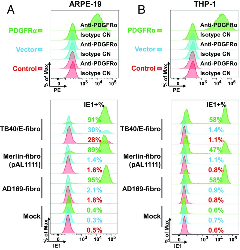
Ectopically expressed PDGFRα renders ARPE-19 and THP-1 cells susceptible to trimer-only virus entry. Cell-surface staining of PDGFRα on ARPE-19 (A) or THP-1 (B) cells following no treatment (control), infection with a lentivirus lacking an insert (vector), or infection with a lentivirus expressing PDGFRα. (Top) Cells were subjected to flow cytometry using the indicated antibodies. (Bottom) Cells were infected with AD169-fibro, Merlin-fibro (pAL1111), or TB40/E-fibro at a multiplicity of 3 FFU per cell, and IE1 expression was measured at 24 hpi (ARPE-19) or 18 hpi (THP-1) to assay the percentage of infected cells.

