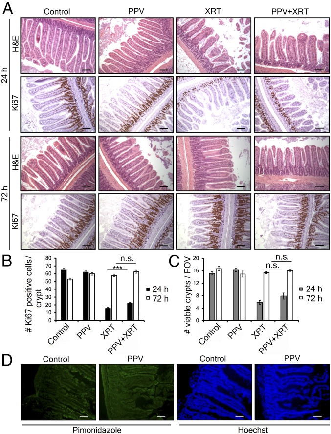
Fig. 3.
Papaverine does not increase radiation-induced normal tissue toxicity. (A) Representative H&E- and Ki67-stained sections of jejunum harvested 24 or 72 h after treatment with papaverine and/or radiation as indicated. (Scale bar, 100 μm.) (B) Quantification of proliferating cells/crypt by Ki67 staining. Data represent mean of at least 30 counted crypts per group (n = 3) ±SEM. (C) The number of regenerating crypts/field of view (FOV). Values represent the mean number of crypts with >10 Ki67-positive cells per FOV ±SEM. P values were calculated with t test. ***P < 0.001; n.s., not significant. (D) Immunofluorescence showing hypoxic marker pimonidazole (green) and Hoechst nuclear counterstain (blue) in jejunum cryosections from MiaPaca-2 tumor-bearing mice treated with saline or 2 mg/kg PPV (n = 4). (Scale bar, 50 μm.)

