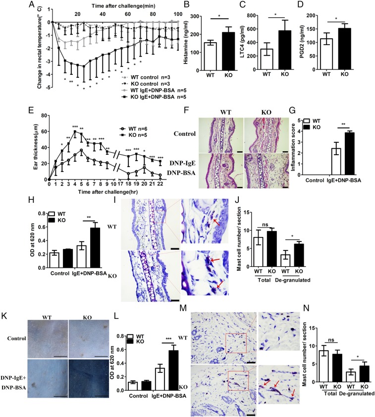
Fig. 4.
RKIP deficiency aggravates PSA and PCA in vivo. (A) Mice were injected i.v. with 10 μg of anti–DNP-IgE per mouse and challenged 24 h later with PBS or 100 μg of DNP-BSA per mouse. Rectal temperatures were assessed every 5 min. (B–D) Releases of (B) histamine, (C) LTC, and (D) PGD2 in the serum were measured at 100 min after DNP-BSA activation. (E) Mice were presensitized by intradermal injection of 3 μg of anti–DNP-IgE per ear (left ear) or an identical volume of PBS (right ear), and, 24 h later, the mice were challenged i.v. with 100 μg of DNP-BSA per mouse in PBS/Evans blue. Ear swelling was calculated as the difference between the thickness of the right and left ears (WT n = 6, KO n = 5). (F) H&E staining and (G) inflammatory cell infiltration scores of the histological sections of the right (control) and left ears (DNP-BSA) from mice treated as in E. (Scale bars in F, 50 μm.) (H) Evans blue dye was extracted in formaldehyde from the reaction sites in E and quantified according to the absorbance at 620 nm. (I) Toluidine blue staining and (J) quantification (blind analysis) of mast cells per section of the left ears from mice treated as shown in E. Red arrows indicate degranulated mast cells. (Scale bars in I, 50 μm.) (K) Mice were presensitized intradermally with anti–DNP-IgE or PBS; 2 d later, the mice were challenged with DNP-BSA in PBS/Evans blue, and the skin was examined visually (n = 6). (Scale bars, 10 mm.) (L) Evans blue dye was extracted in formaldehyde from the reaction sites in K and quantified as absorbance at 620 nm. (M) Toluidine blue staining and (N) quantification (blind analysis) of mast cells per section of back skin (DNP-BSA) from mice treated as in K. Red arrows indicate degranulated mast cells. (Scale bars in M, 50 μm.) ns, no significant difference; *P < 0.05; **P < 0.01; ***P < 0.001. Data are representative of three experiments. Data represent mean and SD.

