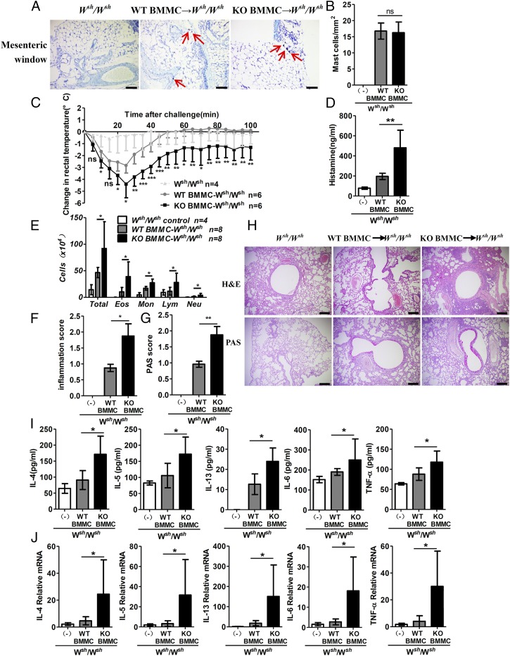
Fig. 5.
RKIP deficiency in mast cells aggravates IgE-mediated allergic response. Mast cell-deficient mice were given no BMMCs (Wsh/Wsh control; n = 4) or were reconstituted with WT BMMCs (WT BMMC → Wsh/Wsh; n = 6) or RKIP KO BMMCs (KO BMMC → Wsh/Wsh; n = 6). (A) Four months later, tissue mast cells in a mesenteric window were assessed in the indicated chimeric mice by toluidine blue staining. Red arrows indicate mast cells. (Scale bars, 50 μm.) (B) Absolute numbers of mast cells per square millimeter in Wsh/Wsh control mice (n = 4) or chimeric mice (n = 6 per group). Reconstituted mice were presensitized with anti–DNP-IgE overnight and challenged 24 h later with 100 μg of DNP-BSA or PBS in Evans blue dye. (C) Rectal temperatures were assessed every 5 min. (D) Histamine release in serum was measured at 100 min after PBS or DNP-BSA challenge. Wsh/Wsh control (n = 5), WT BMMC → W sh/Wsh mice (n = 8), and KO BMMC → Wsh/Wsh mice (n = 8) were challenged with OVA or PBS in the airway inflammation model and analyzed 24 h after the final challenge. (E) Numbers of total cells (total), eosinophils (Eos), neutrophils (Neu), lymphocytes (Lym), and macrophages (Mac) in the BALF were assessed after Wright−Giemsa staining. (F) Inflammation scores and (G) PAS scores were analyzed as described in Materials and Methods. (H) Sections of lung tissues were stained with H&E and PAS staining. (Scale bars, 200 µm.) (I) The mRNA expression of IL-4, IL-5, IL-13, IL-6, and TNF-α in lung tissue was assessed by real-time PCR (n = 6). (J) Secretion of IL-4, IL-5, IL-13, IL-6, and TNF-α in BALF as measured by ELISA (n = 6). ns, no significant difference; *P < 0.05; **P < 0.01. Data are representative of three experiments. Data represent the mean and SD.

