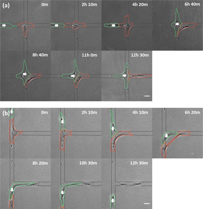
Fig. 1.
Migration of stalled NRK-52E cells after contacting the head of an approaching cell. Phase-contrast images of a cell stalled at an X (A; red outline) or T (B; red outline) intersection show the initiation of migration after interacting with the head of an approaching cell (green outlines). White arrows indicate the direction of migration of the approaching cell. Substrate micropattern is indicated by gray lines. Note both cells on the T micropattern break into two pieces, forming a pair of anuclear cytoplasts along the horizontal branch. Elapsed times are shown above each panel. (Scale bars, 50 µm.)

