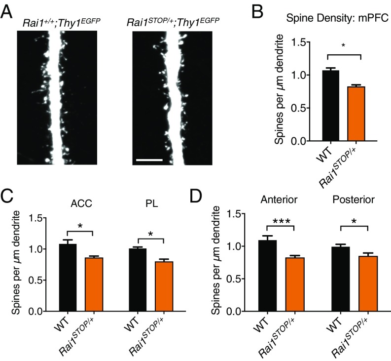
Fig. 4.
Decreased dendritic spine density of mPFC neurons in Rai1STOP/+ mice. (A) Representative image of WT (Rai1+/+;Thy1EGFP) and Rai1STOP/+;Thy1EGFP main apical dendritic trunk of layer V pyramidal neurons at layers II/III. (Scale bar, 5 μm.) (B) Rai1STOP/+ mice (three mice, n = 124 segments) showed a decreased dendritic spine density in the mPFC compared with their WT littermates (three mice, n = 128 segments; mean ± SEM; *P < 0.05; t test). (C) Rai1STOP/+ mice showed decreased spine density in both ACC (WT, n = 71 segments; Rai1STOP/+, n = 68 segments) and PL (WT, n = 53 segments; Rai1STOP/+, n = 60 segments) compared with their WT littermates (mean ± SEM; *P < 0.05; t test). (D) Rai1STOP/+ mice showed decreased spine density in both anterior mPFC (WT, n = 60 segments; Rai1STOP/+, n = 56 segments) and posterior mPFC (WT, n = 64 segments; Rai1STOP/+, n = 72 segments) compared with their WT littermates (mean ± SEM; *P < 0.05, ***P < 0.001; t test). Anterior mPFC showed a greater loss of spine density.

