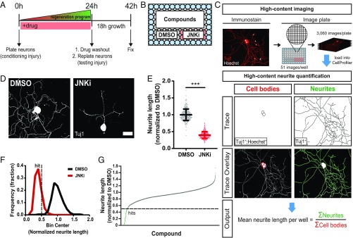
Fig. 1.
A loss-of-function screen identifies inhibitors of axon regeneration signaling in vitro. (A) Primary adult DRG neurons were harvested and dissociated to activate axon injury signaling (conditioning injury). Immediately after plating into 96-well plates, neurons were treated with compounds. After 24 h, the state of the regeneration program was assessed via a replating (testing injury), and neurons were given 18 h to regrow neurites in the absence of drug. (B) Each plate consisted of 60 unique compound wells, 10 wells of the positive control JNKi, and 10 wells of the negative control DMSO. Water (blue) filled the edges of the plate to reduce well-to-well variability. (C) Fixed plates were stained for Hoechst and neuronal Tuj1 and imaged on a high-throughput microscope. For each well, total neurite length per cell was quantified using a custom neurite tracing pipeline built in CellProfiler. Within each well, neurite lengths were summed and divided by the total cell count of the well. (D) Representative images of screen controls. (Scale bar: 50 µm.) (E) Combined data from all control wells in the entire screen: mean ± SD, n = 238–240 for each group, unpaired two-tailed t test, t = 47.1, df = 476, ***P < 0.0001. (F) Histogram of E with hit cutoff at 0.5 (dotted line). (G) Results from screening the ICCB Known Bioactives Library at two doses. Hits are below 0.5 (dotted line).

