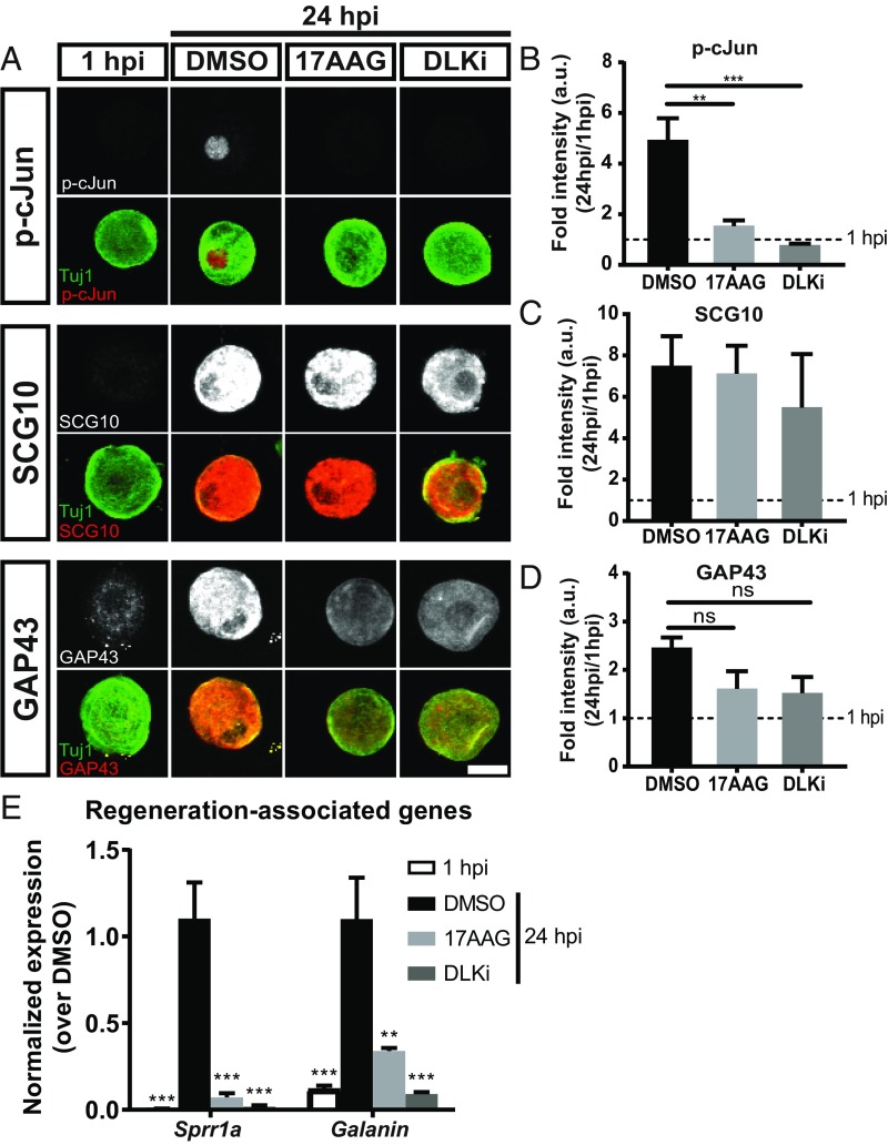
Fig. 3.
Inhibition of HSP90 blocks molecular components of the axon regeneration program. (A) Adult DRG neurons plated and treated with DMSO, 1 µM 17AAG, and 500 nM DLKi. At 24 h postinjury (hpi), adult DRG neurons were fixed and immunostained for proregenerative markers (gray in Top and red in merged) and neuronal Tuj1 (green). (Scale bar: 20 µm.) (B–D) Quantification for each marker (mean ± SEM). Fold intensity was normalized to neurons at 1 hpi (“uninjured”): n = 3–5 independent experiments with ∼40 neurons quantified per group per experiment, one-way ANOVA with Tukey’s multiple comparison test. (B) DF = 17, F = 15.3, DMSO vs. 1 hpi P = 0.0002. **P = 0.001 DMSO vs. 17AAG; ***P = 0.0006 DMSO vs. DLKi. (C) DF = 14, F = 6.64, DMSO vs. 1 hpi P = 0.01, DMSO vs. 17AAG P = 0.99, DMSO vs. DLKi P = 0.80. (D) DF = 14, F = 7.15, DMSO vs. 1 hpi P = 0.004, DMSO vs. 17AAG P = 0.099, DMSO vs. DLKi P = 0.09. (E) Adult DRG neurons were dissociated, plated, and treated with 1 µM 17AAG, 500 nM DLKi, or DMSO. At 24 hpi, RNA was collected, and RAGs were analyzed via qRT-PCR. Fold intensity was normalized to DMSO-treated neurons at 24 hpi: mean ± SEM, n = 5–8 independent experiments, one-way ANOVA with Tukey’s multiple comparison test. For Sprr1a, DF = 26, F = 19.8. ***P < 0.0001 DMSO vs. 1 hpi; ***P < 0.0001 DMSO vs. 17AAG; ***P < 0.0001 DMSO vs. DLKi. For Galanin, DF = 18, F = 14.0. **P = 0.003 DMSO vs. 17AAG; ***P = 0.0005 DMSO vs. 1 hpi; ***P = 0.0002 DMSO vs. DLKi. ns, not significant.

