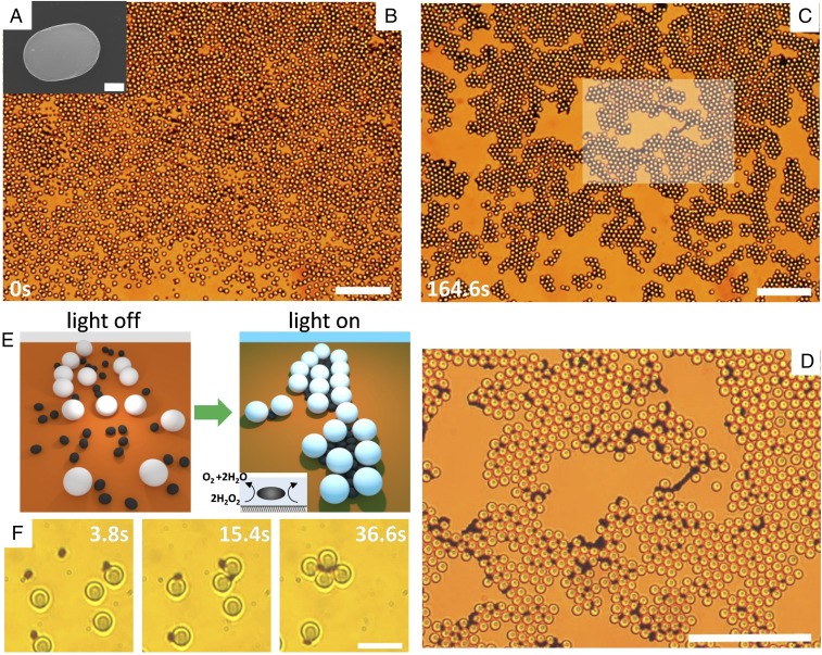
Fig. 1.
(A) Scanning electron microscopy (SEM) image of one hematite ellipsoid (). (Scale bar, .) (B and C) Colloidal gel assembled from doping a bath of silica spheres () with a few hematite ellipsoids in a water solution containing ( vol). The time (C) corresponds to the application of blue light (, ) that triggers the phoretic attraction, and C shows the stationary structure observed after . (D) Enlargement of the central region shown in the box in C. (Scale bars, for all images.) See corresponding video (Movie S1). (E) Schematic showing the assembly of particles due to photoactivated dopants. (F) Formation of one colloidal cluster composed of silica particles, when light is applied at . (Scale bar, .)

